TOOLS
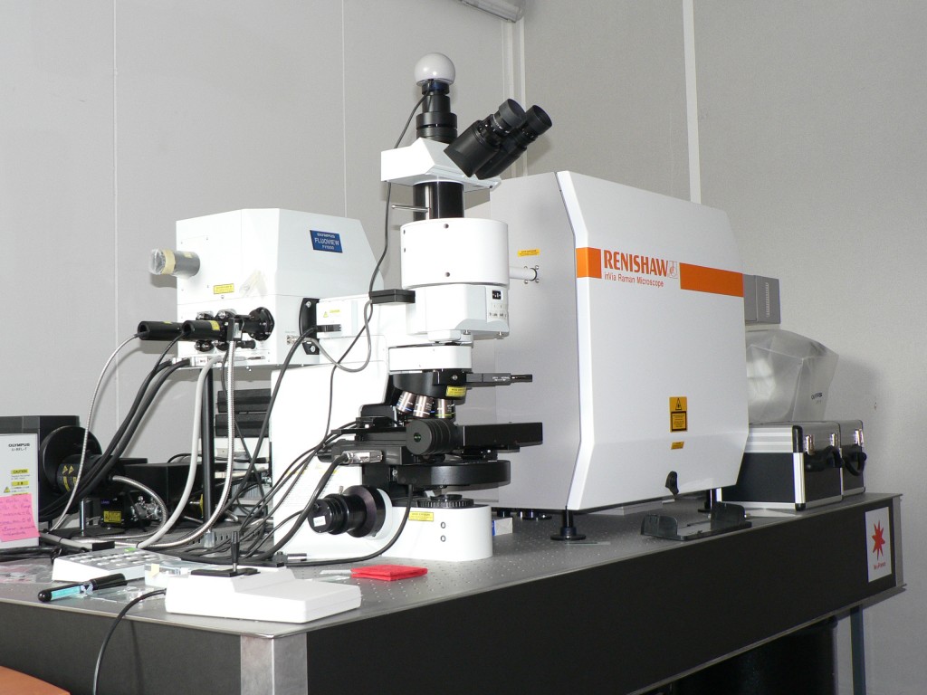
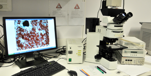
In the recent years, we have used, adapted and improved a variety of techniques to analyse in situ minute amounts of organo-(bio)mineral compounds preserved in rock samples. These include: Raman spectrometer coupled to a confocal fluorescence microscope, scanning electron microscope (SEM), Synchrotron-based techniques (SR-µXRF, SR-µXANES and differential phase contrast imaging techniques) and Secondary Ion Mass Spectrometry (SIMS). Central to these achievements is the technical platform PARI of IPGP and access to large infra-structures such as Synchrotron (SOLEIL, ESRF, SLS, Australian Synchrotron) and Ion Probes (CRPG Nancy, Australian National University) facilities.
THE LAB
Raman/confocal spectroscopy
Optic cathodoluminescence

Optic cathodoluminescence at IPGP, Paris, France. Complete view (left), closer view on the cathode and the vaccum chamber (center) and closer view on the SPOT RT3 camera (right).
Optic microscopy
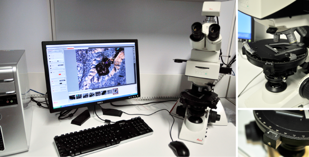
Leica DM2500 P is fully dedicated to petrography.

Transmitted/reflected Leica DM2500 P microscope for petrography research at IPGP, Paris, France. General view (left); detail views on lenses and sample holder (right).
OTHER TECHNIQUES
Synchrotron techniques
Ion microprobes

